Sketch And Label Of A Cross Section Of A Long Bone - Cartilage and Bone | Flickr - Plates of cartilage, also known as growth plates which allow the long bones to grow during childhood.
Sketch And Label Of A Cross Section Of A Long Bone - Cartilage and Bone | Flickr - Plates of cartilage, also known as growth plates which allow the long bones to grow during childhood.. Saved by university of colorado boulder. The structure of a long bone consists of several sections:. Label the haversian canal, osteocyte (mature bone cell) in lacuna, and canaliculi. The hollow region in the diaphysis is called the medullary cavity, which is filled with yellow marrow. Forms the larger rounded ends of long bones.
It is located between the elbow joint and the shoulder. Labeled compact bone microscope slides | labeled histology slides. The walls of the diaphysis are composed of dense and hard compact bone. Use colored pencils to draw and label the following structures as they appear using the 40x objective, or by looking at an image from the internet. Label the membrane that lines the cavity and the membrane that covers the outside surface.
Blood supply of long bones from image.slidesharecdn.com External circumferential lamellae, osteon, central canal, perforating canals, lacuna, canaliculi, concentric lamellae. We start our section on tissue structure function with bone tissue. A long bone has two parts: Figure 5—2b is a drawing of a longitudinal section of the femur. (do not copy and paste a picture from the text or internet.) Related posts of cross section of a long bone bone test anatomy and physiology. This is the long central shaft. The diaphysis and the epiphysis.
Figure 5—2b is a drawing of a longitudinal section of the femur.
Sketch and label a cross section of a bone. The hollow region in the diaphysis is called the medullary cavity, which is filled with yellow marrow. Skeleton system histology slides med lab microscope slides phlebotomy college hacks anatomy and physiology love my job biology. Related posts of cross section of a long bone bone test anatomy and physiology. Labeled compact bone microscope slides | labeled histology slides. Do not color the articular cartilage; Make a pencil sketch and use markers or colored pencils to add details. We start our section on tissue structure function with bone tissue. The inner portion of the bone is composed of trabecular a hand drawn sketch by dr. Osteon, central canal, blood vessels, lamellae, osteocytes, lacunae, canaliculi, and perforating canal. Learners should accurately draw a long bone, resembling that in figure 6.24. The diaphysis and the epiphysis. Exploring the microscopic anatomy of bone 1.
This is the long central shaft. Sketch a longitudinal section through a long bone and label the following structures de epiphysim ercavi periosteum, co p pseen, compact bune.no red bone marrow, and yellow bone marrow he provides a epiphysis riedullary activity 4: Related posts of cross section of a long bone bone test anatomy and physiology. Create a drawing of the bone section in your laboratory journal and label the areas listed above. Do not color the articular cartilage;
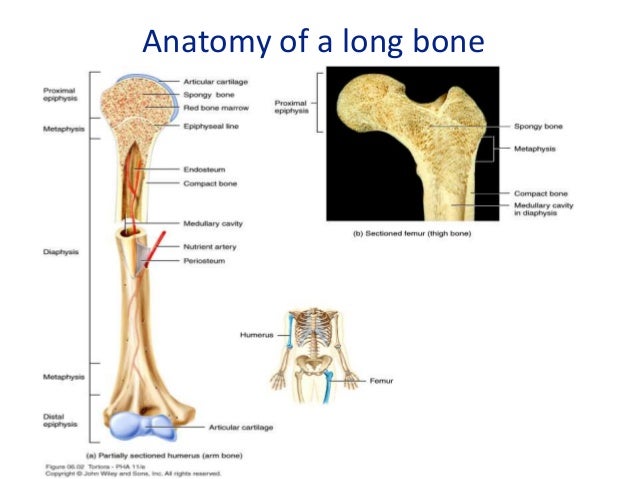
A List of All the Flat Bones in the Human Body With Diagrams from www.buzzle.com In these labeled examples, a human femur is represented without identifying many of the unique characteristics that help differentiate the femur bone from other bones in the human body. The diaphysis and the epiphysis. The structure of a long bone allows for the best visualization of all of the parts of a bone ().a long bone has two parts: It is located between the elbow joint and the shoulder. The diaphysis and the epiphysis. Bone test anatomy and physiology 12 photos of the bone test anatomy and physiology anatomy and physiology bone lab test, anatomy and physiology bone markings test, anatomy and physiology bone practical test, anatomy and physiology bone tissue test, anatomy and physiology test on bone tissue, bone, anatomy and. The structure of a long bone allows for the best visualization of all of the parts of a bone (figure 6.7). The hollow region in the diaphysis is called the medullary cavity, which is filled with yellow marrow.
The diaphysis is the tubular shaft that runs between the proximal and distal ends of the bone.
The humerus is the long bone in the upper arm. At the elbow, it connects primarily to the ulna, as the forearm's radial bone connects to the. The hollow region in the diaphysis is called the medullary cavity, which is filled. The diaphysis and the epiphysis. Long bones have a thick outside layer of compact bone and an inner medullary cavity containing bone marrow. Also known as the middle phalanx, the short pastern bone sits on top of the articulating joint of the pedal bone and underneath the long pastern bone. This is for two reasons: The digital cushion sits just behind the pedal bone and above the sensitive frog. Exploring the microscopic anatomy of bone 1. A long bone is a bone that has greater length than width. Then, fill in the table below to describe each. In these labeled examples, a human femur is represented without identifying many of the unique characteristics that help differentiate the femur bone from other bones in the human body. Labeled compact bone microscope slides | labeled histology slides.
Label the haversian canal, osteocyte (mature bone cell) in lacuna, and canaliculi. Figure 5—2b is a drawing of a longitudinal section of the femur. Make a pencil sketch and use markers or colored pencils to add details. Only the bottom portion of this bone extends as far as the hoof capsule. Once we stop growing (between 18.
Cross section of an artery, vein and capillary | Nursing ... from s-media-cache-ak0.pinimg.com The diaphysis and the epiphysis. Label the membrane that lines the cavity and the membrane that covers the outside surface. A long bone has a shaft and 2 ends. The structure of a long bone allows for the best visualization of all of the parts of a bone (figure 6.7). The head of each end of a long bone consists largely of spongy bone and is covered with hyaline cartilage. The walls of the diaphysis are composed of dense and hard compact bone. Long bones have a thick outside layer of compact bone and an inner medullary cavity containing bone marrow. A long bone has two parts:
The diaphysis is the tubular shaft that runs between the proximal and distal ends of the bone.
The diaphysis and the epiphysis. Smartdraw includes 1000s of professional healthcare and anatomy chart templates that you can modify and make your own. Related posts of cross section of a long bone bone test anatomy and physiology. Sketch a longitudinal section through a long bone and label the following structures de epiphysim ercavi periosteum, co p pseen, compact bune.no red bone marrow, and yellow bone marrow he provides a epiphysis riedullary activity 4: Bone not color the articular cartilage; A long bone has a shaft and 2 ends. A long bone has two parts: Draw and label a longitudinal section of a long bone. Human back muscles and bones 12 photos of the human back muscles and bones human back muscles and bones, bone, human back muscles and bones. On this page, you will find two images i created that illustrate the parts of a long bone and long bone structure. Skeleton system histology slides med lab microscope slides phlebotomy college hacks anatomy and physiology love my job biology. Saved by university of colorado boulder. The diaphysis is the tubular shaft that runs between the proximal and distal ends of the bone.


0 Komentar